Calcaneovalgus/calcaneovarus test
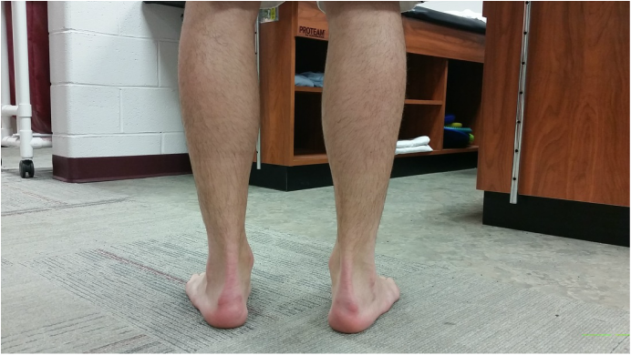
Patient Position
- Have the patient stand on a flat surface such as the floor. You may have the patient stand on a table if it makes it easier to view the calcaneus.
Directions
- View the posterior aspect of the patient's achilles tendon and calcaneus and make note of their position.
- Make note of the number of phalanges (toes) that are visible laterally.
Expected Results
- You should see the achilles tendon extending straight down to the calcaneus. A patient with this anatomical build is considered to have a neutral calcaneus.
- You should be able to view no more than two of the lateral phalanges(the 4th and 5th phalanges).
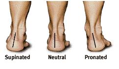
Positive Signs
- The calcaneus protudes either laterally (calcaneovarus or supinated foot) or medially (calcaneovalgus or pronated foot).
- When viewing the patients feet from a posterior aspect there are more than two phalanges visible laterally on either side.
Implications
- A positive sign where the calcaneus protrudes laterally indicates that the patient is supinated (pes planus or more commonly referred to as flat footed).
- A positive sign present where the calcaneus protrudes medially indicates that the patient is pes cavus (high arches).
- A positive sign where there are more than 2 visible phalanges laterally indicates that the patient has an extreme pes planus condition.
- Have the patient stand on a flat surface such as the floor. You may have the patient stand on a table if it makes it easier to view the calcaneus.
Directions
- View the posterior aspect of the patient's achilles tendon and calcaneus and make note of their position.
- Make note of the number of phalanges (toes) that are visible laterally.
Expected Results
- You should see the achilles tendon extending straight down to the calcaneus. A patient with this anatomical build is considered to have a neutral calcaneus.
- You should be able to view no more than two of the lateral phalanges(the 4th and 5th phalanges).
Positive Signs
- The calcaneus protudes either laterally (calcaneovarus or supinated foot) or medially (calcaneovalgus or pronated foot).
- When viewing the patients feet from a posterior aspect there are more than two phalanges visible laterally on either side.
Implications
- A positive sign where the calcaneus protrudes laterally indicates that the patient is supinated (pes planus or more commonly referred to as flat footed).
- A positive sign present where the calcaneus protrudes medially indicates that the patient is pes cavus (high arches).
- A positive sign where there are more than 2 visible phalanges laterally indicates that the patient has an extreme pes planus condition.
|
Supinated foot
- Calcaneovalgus = pes planus = flat footed Nuetral foot - Normal foot Pronated foot - Calcaneovarus = pes cavus = high arches |
Treatment Options
- An indiviual who presents with foot supination can be treated with orthotics or athletic taping. This condition is most effectively treated by using an orthotic with a built in arch to mimic the presence of a normal arch. This condition is commonly treated by athletic trainers by using a basic arch tape job.
- An individual who presents with foot pronation can be treated with the use of orthotics. This condition is most effectively treated with a custom orthotic molded to fit the shape of the patient's foot.
Rehabilitation Direction
- The rehabilitation program for an individual that presents with a supinated foot should include exercises to strengthen the plantar fascia of each foot.
- The rehabilitation program for an individual that presents with a pronated foot should include a stretching routine focused on lengthening the plantar fascia. For extreme cases a night splint can be used to stretch the plantar fascia as the patient sleeps.
|
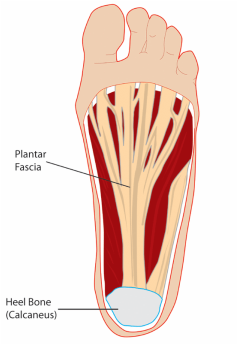
The plantar fascia is located on the bottom of the foot an spans from the calcaneus to the ball of the foot. At the distal end, the planar fascia attaches to the ball of the foot proximal to each of the five phalanges. The length of the plantar fascia is the key determinant to weather or not the patient is supinated or pronated. |
