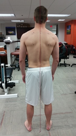
Patient Position
- Have the patient stand with their feet shoulder width apart in a relaxed (normal) position.
- The patient should be close to the plumb line without touching the suspended yarn.
Directions
- Have patient stand close to the plumb line without touching the yarn.
- Assure that the plumb line evenly bisects the patients gluteus maximus.
- Capture an Image of the patient in this position to use for the analysis.
Note
- Some of the data that you could gather from this view was covered in the previous testing areas and will therefore not be part of this area of the analysis.
Key Points of the Analysis
- Sacroilliac Joints - View the SI Joint of the patient and check for the level of each SI Joint. The SI joints are commonly known as the dimples of the lower back.
- Spine - View the shape of the vertebral line and make note of any contour in from the lower back to the cervical vertebrae distal to the base of the skull.
Implications and Rehabilitation Direction
- Sacroilliac Joints - The SI joints should appear parallel when viewed bilaterally. Nonparallel SI joint appearance indicates pelvic misalignment and can be treated with orthopedic manipulative treatment. Patient may need several treatments to permanently change the alignment of their pelvis. This treatment should be administered by certified osteopathic doctors only.
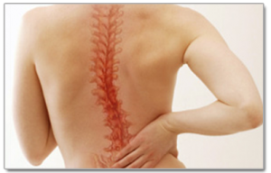
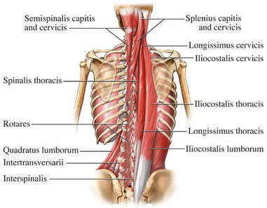
- Spine - The Spine should appear straight and run evenly with the visible plumb line. Any contour in the vertebral line may indicate either muscular weakness (covered in the scapular position test) or mild to severe scoliosis. Scoliosis is a condition where there is a sideways curvature present in the spine of an individual. Consult a physician for treatment options of severe scoliosis as this condition usually requires the uses of an orthopedic back brace. For patients who display mild to moderate scoliosis you can treat with prescription rehabilitation exercises focusing primarily on the core muscles and the intraspinal muscles. The intraspinal muscles are a large group of muscles that are shown in the graphic below (right) and are responsible for spinal stabilization.
- Sacroilliac Joints - The SI joints should appear parallel when viewed bilaterally. Nonparallel SI joint appearance indicates pelvic misalignment and can be treated with orthopedic manipulative treatment. Patient may need several treatments to permanently change the alignment of their pelvis. This treatment should be administered by certified osteopathic doctors only.
- Spine - The Spine should appear straight and run evenly with the visible plumb line. Any contour in the vertebral line may indicate either muscular weakness (covered in the scapular position test) or mild to severe scoliosis. Scoliosis is a condition where there is a sideways curvature present in the spine of an individual. Consult a physician for treatment options of severe scoliosis as this condition usually requires the uses of an orthopedic back brace. For patients who display mild to moderate scoliosis you can treat with prescription rehabilitation exercises focusing primarily on the core muscles and the intraspinal muscles. The intraspinal muscles are a large group of muscles that are shown in the graphic below (right) and are responsible for spinal stabilization.
|
|
